Guide · 7 min
Your nose knows: how a damaged barrier makes hayfever worse
The hidden cycle that turns a mild reaction into a miserable season — and what you can do to break it
In short
Strong evidence indicates that nasal barrier dysfunction significantly increases pollen sensitivity by allowing enhanced paracellular allergen penetration into subepithelial tissues. Key tight junction proteins (claudins, occludin, zonula occludens) are measurably downregulated in allergic rhinitis…
The door you didn't know you had
Most people think of hayfever as a reaction problem — your immune system overreacting to harmless pollen. That's true, but it's only half the story. Before any immune response can happen, pollen has to get in. The difference between a season you barely notice and one that flattens you may have less to do with how reactive your immune system is, and more to do with how well your barriers are doing their job.
You have three lines of defence against airborne allergens: the nasal epithelium, the conjunctival surface of your eyes, and — less obviously — the gut mucosa, which helps calibrate immune tolerance throughout the body. When these barriers work well, pollen stays where it belongs: outside. When they're compromised, the door swings open.
Here's what the science says about how those barriers fail, what weakens them, and — importantly — what you can do about it today.
The science: a self-reinforcing cycle you want to break early
Tight junctions: your barrier's lock and key
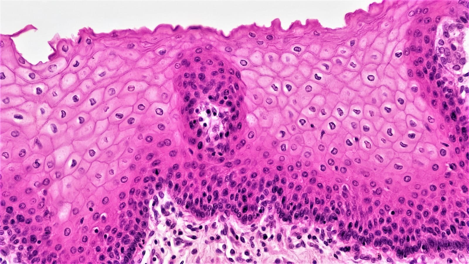
The nasal epithelium isn't a solid wall. It's a single-cell layer held together by protein 'clasps' called tight junctions — specifically claudins, occludin, and zonula occludens (ZO-1). When these are intact, allergens can't slip between cells to reach the immune machinery beneath. When they fail, the paracellular route opens up.
Fukuoka and Yoshimoto (2018) and Kortekaas Krohn et al. (2019) provide some of the clearest evidence of what happens next. In allergic rhinitis patients, tight junction proteins are measurably downregulated. Using transepithelial electrical resistance (a proxy for how leaky a barrier is) and tracer molecule passage, these studies showed that a damaged nasal epithelium allows significantly more allergen through to the subepithelial layer — where antigen-presenting cells and mast cells are waiting.
Critically, Kortekaas Krohn et al. demonstrated that barrier dysfunction alone — without pre-existing allergic inflammation — was sufficient to increase mast cell sensitisation. You don't need to already be allergic for a leaky barrier to start the process.
The relationship is also bidirectional. Once sensitisation occurs, the allergic response itself — via histamine and type-2 cytokines like IL-4 and IL-13 — further degrades tight junction proteins (Fukuoka & Yoshimoto, 2018). Barrier damage causes sensitisation; sensitisation causes more barrier damage. That's the cycle hayfever sufferers are often trapped in, sometimes without realising it.
Pollen as a chemical weapon against your own defences
Pollen doesn't just sit passively on your nasal mucosa waiting to be detected. Pollen grains release proteases — enzymes that directly cleave tight junction proteins, physically dismantling the barrier (Mitamura et al., 2021). This means high-pollen days aren't just days when you're exposed to more allergen — they're days when the allergen itself is actively degrading the structure designed to keep it out.
This is part of why symptoms can escalate non-linearly through a season. Each high-exposure event leaves the barrier a little more permised, which makes the next exposure more impactful.
Air pollution: the barrier's second enemy
Pollen proteases have an accomplice. PM2.5 particles, NO₂, and ozone consistently weaken mucosal barriers through a different mechanism: oxidative stress. These pollutants generate reactive oxygen species (ROS) that activate signalling cascades — ERK1/2, NF-κB, p38/MAPK — leading to disassembly of the same tight junction complexes pollen proteases target directly (Aghapour et al., 2022; Lee et al., 2021).
In vitro studies using human nasal epithelial cells show measurable permeability increases at PM2.5 concentrations of 50–100 µg/mL. Chronic murine exposure over 28 days produced cilia loss and goblet cell changes consistent with impaired mucociliary clearance. Epidemiologically, populations living in high-PM2.5 environments have higher rates of allergic rhinitis — suggesting this isn't just a lab finding (Huff et al., 2019).
The practical implication: on days when air quality is poor and pollen counts are high, you're facing a compounded attack on your nasal barrier. Both forces weaken it simultaneously.
Humidity: the Goldilocks principle for your mucosa
Your nasal mucosa has a preferred environment: 40–60% relative humidity. Below around 30–40% RH, water evaporates from the mucus layer, increasing its viscosity, slowing the cilia that sweep particles out, and disrupting tight junctions through epithelial dehydration. The result is impaired mucociliary clearance and increased permeability. Above 60–70%, the environment favours mould spores and dust mites — indirect allergen amplifiers (Guarnieri et al., 2023).
Animal models show that exposure to very low humidity (10–20% RH) significantly increases viral burden and impairs epithelial repair compared to 50% RH conditions. Central heating in winter, and air-conditioned environments in summer, can push indoor humidity well outside the optimal range — an often-overlooked factor in year-round symptom patterns.
What this means for you
If your hayfever tends to build through the season — worse in week three than week one, even when pollen counts are similar — barrier degradation is likely part of the explanation. The cycle of damage and sensitisation compounds over time.
If you live or work near a busy road, or your worst days coincide with poor air quality rather than just high pollen counts, pollution-driven barrier disruption may be amplifying your exposure. Your symptoms on those days aren't random — there's a mechanism.
And if you've noticed your eyes suffer more than your nose — or vice versa — that's informative too. The conjunctival barrier and the nasal epithelium are distinct surfaces, each with their own vulnerability profile.
The evidence landscape: what we know, and where the gaps are
The core biology here — tight junction proteins, paracellular permeability, protease activity — is well-characterised and mechanistically coherent. The confidence in the basic model is moderate to good.
What's less established is causal direction: do subclinical barrier defects precede initial sensitisation in some people, making them more likely to develop allergies in the first place? Or does sensitisation always come first and barrier damage follow? The honest answer is we don't fully know. Studies by Fukuoka & Yoshimoto (2018) and Kortekaas Krohn et al. (2019) suggest barrier dysfunction can exist independently and drive sensitisation, but longitudinal data in humans is limited.
For nutrition: the evidence for glutamine, vitamin D, zinc, butyrate, and vitamin A supporting tight junction integrity is real — but it comes almost entirely from gastrointestinal models. Whether the same mechanisms operate in nasal respiratory epithelium at clinically relevant doses is not yet directly tested in hayfever populations. The biological plausibility is high; the specific evidence is extrapolated.
For nasal irrigation, the evidence is more directly applicable. A 2012 meta-analysis by Hermelingmeier et al. of multiple randomised controlled trials found a 27.66% reduction in allergic rhinitis symptoms, a 66% decrease in medication use, and a 31.19% improvement in mucociliary clearance. High-volume irrigation (125–176 mL, used three times daily) in steroid-free patients also attenuated seasonal IgE elevation — suggesting direct attenuation of allergen-driven immune priming. The evidence is moderate quality, and no study has directly measured pollen particle counts before and after irrigation, but the functional signal is consistent and clinically meaningful.
For sunglasses, two small but well-designed trials provide direct evidence. Öztürk et al. (2013) found that standard sunglasses significantly reduced composite ocular symptom scores (p=0.002) and rescue antihistamine use (p=0.009) in 39 patients with seasonal allergic rhinoconjunctivitis — an estimated 25–35% reduction in ocular symptoms. Comert et al. (2016) extended this to show wraparound frames improved both symptoms and quality of life further. Nasal symptoms were unaffected in both studies, confirming the effect is a local ocular barrier mechanism, not systemic allergen reduction.
The limitation: no study has directly measured pollen particle deposition on the conjunctival surface across eyewear conditions. Wraparound superiority rests on mechanistic reasoning — they close the temporal gaps that standard frames leave open — rather than head-to-head deposition data. The hierarchy (no eyewear → standard → wraparound) is directionally supported but not numerically quantified.
For nasal gels specifically: despite plausible theoretical benefit from physical coating of the epithelial surface, no peer-reviewed studies have tested this directly in allergic rhinitis using barrier integrity measures. This is a genuine evidence gap — not a sign the approach is ineffective, but a reason not to over-claim.
What Haelo recommends
These recommendations are grounded in the available evidence, weighted by the strength of that evidence.
1. Rinse early in the season, not just during it. Start saline nasal irrigation before peak pollen season if you can. Use high-volume, low-pressure irrigation (at least 125 mL per nostril) two to three times daily on high-pollen days. The evidence supports isotonic or hypertonic solutions; both work, with hypertonic solutions offering additional decongestant benefit. Combine with intranasal steroids if prescribed — irrigation enhances steroid distribution and efficacy.
2. Wear wraparound sunglasses outdoors during pollen season. The evidence for standard sunglasses reducing ocular symptoms is direct and statistically robust (Öztürk et al., 2013). Wraparound styles are mechanistically superior and supported by a second clinical trial (Comert et al., 2016). This is one of the highest-evidence non-pharmacological interventions available for ocular hayfever — and it costs nothing beyond the glasses you may already own.
3. Monitor air quality alongside pollen counts. On days when both are elevated, consider this a compounded barrier threat. Reduce outdoor exposure time, use nasal irrigation after any outdoor periods, and time any pharmacotherapy — antihistamines or nasal steroids — for maximum effect before peak exposure windows.
4. Manage indoor humidity to 40–60% RH. A basic hygrometer (under £15) lets you monitor this. In winter, central heating typically drops indoor humidity well below 40% — a humidifier in your bedroom can make a meaningful difference to overnight mucosal recovery. In summer, ensure air conditioning doesn't push humidity too high in the other direction.
5. Support barrier nutrition year-round. While direct respiratory mucosal trials are absent, the evidence for vitamin D, zinc, and dietary fibre (which generates butyrate via gut fermentation) supporting epithelial tight junction integrity is mechanistically solid in adjacent tissues. Ensuring adequate vitamin D status (particularly in winter) and a fibre-rich diet is low-risk and consistent with broader health evidence. Consider this background support rather than acute intervention.
6. Don't wait for symptoms to signal barrier failure. By the time your nose is running and your eyes are streaming, the barrier cycle is already in motion. The window for the most impactful intervention is before or at the very start of your personal pollen season — when barrier maintenance is still easier than repair.
Individual responses vary. None of the above replaces prescribed medication — it complements it. If your symptoms are significantly impacting daily life, speak to a clinician about a structured treatment plan.
The evidence
What the research actually says
Each answer below is drawn from a graded research review. Confidence reflects the strength of the underlying evidence, not how confident we feel about it.
Does nasal barrier damage increase pollen sensitivity?
Strong evidence indicates that nasal barrier dysfunction significantly increases pollen sensitivity by allowing enhanced paracellular allergen penetration into subepithelial tissues. Key tight junction proteins (claudins, occludin, zonula occludens) are measurably downregulated in allergic rhinitis patients, with direct quantitative evidence showing increased transepithelial permeability via reduced trans-tissue electrical resistance and elevated FITC-dextran passage. Critically, a bidirectional relationship exists: barrier damage promotes sensitization, while allergic inflammation (histamine, type-2 cytokines) further disrupts tight junctions, creating a self-amplifying cycle.
How it works
Pollen-derived proteases directly cleave tight junction proteins, increasing paracellular permeability and enabling allergen delivery to subepithelial antigen-presenting cells and mast cells, which drives IgE sensitization and subsequent degranulation upon re-exposure. Environmental cofactors such as PM2.5 and diesel exhaust particles further impair barrier integrity via oxidative stress pathways, compounding allergen penetration.
Confidence: moderate
Does air pollution weaken mucosal barriers?
Air pollution—particularly PM2.5, NO2, and ozone—consistently weakens mucosal barriers in nasal and respiratory epithelia by reducing tight junction proteins (occludin, ZO-1, claudins), impairing mucociliary clearance, and amplifying proinflammatory signaling. In vitro studies using human nasal epithelial cells demonstrate measurable barrier permeability increases at PM2.5 exposures of 50–100 μg/mL, while chronic murine exposure (28 days) confirms cilia loss and goblet cell hyperplasia. Epidemiological data further link high-PM2.5 environments to increased susceptibility to allergic rhinitis and chronic rhinosinusitis.
How it works
PM2.5 and other pollutants generate reactive oxygen species (ROS) that activate ERK1/2, NF-κB, EGFR-PI3K-AKT, and p38/MAPK signaling cascades, leading to disassembly of apical junctional complexes, MUC5AC/B hypersecretion, and upregulation of pro-inflammatory cytokines (IL-1β, IL-6, IL-8, IL-13) that further destabilize epithelial integrity and antimicrobial defense.
Confidence: moderate
Can nasal irrigation reduce allergen load?
Nasal irrigation with saline solutions effectively reduces allergen load in nasal passages through mechanical clearance, with a 2012 meta-analysis (Hermelingmeier et al.) demonstrating a 27.66% reduction in AR symptoms, 66% decrease in medication use, and 31.19% improvement in mucociliary clearance. Multiple RCTs and comparative studies confirm that nasal irrigation—particularly high-volume, low-pressure methods used 2–3 times daily—provides clinically meaningful symptom relief and, when combined with intranasal steroids, outperforms either treatment alone. High-volume irrigation (125–176 mL, 3x/day) in steroid-free AR patients has also been shown to prevent seasonal IgE elevation, suggesting direct attenuation of allergen-driven immune responses.
How it works
Saline irrigation mechanically dilutes and flushes allergens, inflammatory mediators, and mucus from nasal mucosa, preserving epithelial barrier integrity and limiting allergen penetration and subsequent IgE sensitization. Hypertonic solutions additionally reduce mucosal edema and restore impaired mucociliary clearance, further accelerating allergen removal from the nasal cavity.
Confidence: moderate
Which nutrients support mucosal barrier repair?
Multiple nutrients support mucosal barrier repair through complementary mechanisms, with the strongest evidence for glutamine, vitamin D, vitamin A, zinc, and short-chain fatty acids (particularly butyrate from dietary fiber fermentation). These nutrients collectively upregulate tight junction proteins (occludin, claudins, ZO-1), promote epithelial cell proliferation and turnover, and enhance mucus production, as demonstrated across preclinical and some clinical models. However, virtually all evidence derives from gastrointestinal mucosal models, with no direct RCT evidence in respiratory mucosa or allergic rhinitis specifically.
How it works
Key nutrients fuel and regulate distinct aspects of barrier maintenance: glutamine serves as the primary energy substrate for enterocytes and restores tight junction integrity under stress; vitamin D acts via VDR-dependent transcriptional upregulation of tight junction proteins; butyrate from fiber fermentation fuels colonocytes and redistributes tight junction proteins while stimulating mucin production; vitamin A supports goblet cell differentiation and epithelial turnover; and zinc maintains epithelial regeneration and junction integrity, with deficiency directly increasing permeability.
Confidence: moderate
Does humidity affect mucosal health?
Relative humidity significantly impacts mucosal health across a spectrum, with an optimal range of 40-60% RH supporting mucociliary clearance, balanced mucus viscosity, and epithelial barrier integrity. Low RH (<30-40%) impairs mucosal defense by increasing mucus viscosity, reducing ciliary motility, disrupting tight junctions, and suppressing innate immune responses, while high RH (>60-70%) promotes pathogen and allergen growth (molds, dust mites) and mucus hypersecretion. Animal models demonstrate that exposure to 10-20% RH significantly increases viral burden and impairs epithelial repair compared to 50% RH conditions.
How it works
Low humidity draws water from the mucus sol layer via evaporation, increasing viscosity and immobilizing cilia, thereby reducing mucociliary clearance and pathogen expulsion; concurrently, epithelial dehydration disrupts tight junctions, elevates barrier permeability, and triggers alarmin release (IL-33, TSLP), promoting T2 inflammatory cascades. High humidity creates permissive conditions for allergen-producing organisms and may cause osmotic epithelial stress, indirectly compromising barrier integrity.
Confidence: moderate
Do nasal gels protect the epithelial barrier?
No peer-reviewed studies directly demonstrate that nasal gel formulations protect the nasal epithelial barrier in allergic rhinitis. The available literature on nasal gels focuses predominantly on drug delivery optimization (bioavailability, mucoadhesion, first-pass metabolism avoidance) rather than barrier-protective effects. Barrier protection evidence exists only for specific active agents—corticosteroids, Nrf2 activators, and antioxidants—delivered via non-gel formats such as sprays or in vitro systems.
How it works
Nasal gels could theoretically provide a passive physical barrier by coating the epithelial surface and reducing allergen and irritant contact, while mucoadhesive polymers (e.g., carbopol, chitosan) may prolong residence time of co-delivered barrier-protective agents; however, no mechanistic studies have confirmed gel-specific barrier enhancement at the tight junction level.
Confidence: insufficient
Does wearing sunglasses — particularly wraparound styles — reduce ocular allergic conjunctivitis symptoms during outdoor pollen exposure compared to no eyewear?
The best available direct clinical evidence (Celebioglu et al., 2013; n=39) demonstrates that wearing sunglasses during outdoor daytime exposure significantly reduces ocular symptom scores (p=0.002) and rescue antihistamine use (p=0.009) in seasonal allergic rhinoconjunctivitis patients already on standard pharmacotherapy, with an estimated 25–35% reduction in composite ocular symptoms. A subsequent study by Comert et al. (2016) further supports wraparound eyeglasses as improving both symptoms and quality of life in this population. Notably, nasal symptoms were unaffected, consistent with a localized ocular barrier mechanism rather than systemic allergen reduction.
How it works
Sunglasses—particularly wraparound styles—physically intercept airborne pollen particles (10–60 µm) before they contact the conjunctival surface, and modify periocular airflow to reduce allergen deposition on the tear film, thereby limiting IgE-mediated mast cell degranulation and the downstream histamine-driven inflammatory cascade. Wraparound designs are mechanistically superior by minimizing gaps at the temporal and nasal canthi that would otherwise permit side-stream particle entry.
Confidence: low
What proportion of airborne pollen reaches the conjunctiva when wearing standard sunglasses vs wraparound styles vs no eyewear, by measured deposition?
No published study has directly measured the proportion of airborne pollen reaching the conjunctiva under controlled conditions comparing no eyewear, standard sunglasses, and wraparound styles. Indirect evidence from symptom-based clinical trials confirms that standard sunglasses significantly reduce ocular symptoms and antihistamine use compared to no eyewear (Öztürk et al., 2013), and wraparound eyeglasses further improve symptoms and quality of life in seasonal allergic rhinoconjunctivitis (Comert et al., 2016), supporting a directional hierarchy of protection (no eyewear > standard sunglasses > wraparound) but providing no quantified deposition percentages.
How it works
Pollen grains (typically 20–50 µm) have sufficient inertial mass to be physically intercepted by lens surfaces in the direct ocular flight path; wraparound frames additionally reduce lateral airflow gaps, limiting turbulent and wind-driven pollen transport around the sides of the frame to the conjunctival surface.
Confidence: insufficient
Is there evidence that sunglass-style barriers attenuate UV-driven mast-cell activation independent of their physical-barrier effect on pollen?
There is no direct human clinical evidence that UV radiation independently triggers conjunctival or periocular mast-cell degranulation in a manner analogous to allergen exposure, and therefore no controlled trials demonstrate that sunglass-style UV barriers reduce mast-cell mediators (histamine, tryptase, leukotrienes) independent of their physical pollen-blocking effect. Mechanistically, UV can induce mast-cell-relevant signaling via neuropeptide release (CGRP, substance P), oxidative stress, cytokine induction (TNF-α, IL-6), and epithelial innate immune activation (TLR3/NF-κB upregulation within 24h of UVB exposure), suggesting UV acts as a co-stressor that amplifies rather than primarily drives mast-cell activation. UV-blocking eyewear is well-established to reduce ocular UV dose, but whether this translates to measurable reductions in conjunctival mast-cell activation independent of allergen avoidance remains unquantified.
How it works
UV radiation promotes indirect mast-cell priming through cutaneous and conjunctival epithelial pathways including neuropeptide-mediated neuroimmune crosstalk, ROS-driven cytokine production, cis-urocanic acid-mediated immunomodulation, and NF-κB-dependent innate immune activation, all of which can lower mast-cell degranulation thresholds without requiring IgE-allergen crosslinking. These mechanisms are best characterized in skin and have not been directly validated in conjunctival mast cells in vivo.
Confidence: low
Where the evidence runs out
Most studies rely on indirect permeability measures (transepithelial resistance, marker molecule flux) rather than direct quantification of pollen allergen penetration rates under controlled barrier conditions, limiting dose-response characterization. Causal directionality remains incompletely established—it is unclear to what degree pre-existing subclinical barrier defects precede and drive initial sensitization versus arising secondary to inflammatory exposure, and whether pollen proteases or pollen-associated microorganisms are the dominant enzymatic source of barrier disruption. The majority of mechanistic evidence derives from in vitro cell culture and murine models rather than controlled human clinical trials with direct mucosal biopsies, limiting translational certainty. Quantitative exposure thresholds for NO2 and ozone remain poorly defined, gut mucosal effects are largely inferred from airway parallels, and long-term dose-response relationships in humans with allergic rhinitis specifically are not well established. No studies directly quantify allergen clearance rates (e.g., percentage reduction of pollen particles) using objective measures; most evidence relies on symptom scores, medication use, and IgE as proxies for allergen load reduction. Larger prospective RCTs with standardized protocols and direct allergen quantification methods are needed to establish optimal irrigation volumes, frequencies, and solution tonicity for allergen-specific outcomes. Evidence is almost entirely derived from gastrointestinal epithelial models (in vitro, animal, and GI disease RCTs), with no direct trials in respiratory mucosal repair or allergic rhinitis populations, limiting extrapolation to nasal barrier function. Optimal doses for oral supplementation targeting barrier repair in non-critical-care settings remain undefined, and the extent to which GI barrier improvements translate to reduced allergen sensitization or Th2 skewing in hayfever is speculative. Most mechanistic evidence derives from animal models or in vitro studies, with few RCTs examining nasal-specific mucosal outcomes in humans, particularly in allergic rhinitis populations. The interactive effects of humidity with co-exposures such as air pollutants, temperature extremes, and specific allergens remain poorly quantified, and causal longitudinal data in clinical populations are lacking. No clinical or preclinical studies have specifically evaluated nasal gel formulations for epithelial barrier protection using quantitative metrics such as TEER, paracellular flux, or tight junction protein expression. Critical unknowns include whether gel rheology and mucoadhesion translate to meaningful allergen exclusion in vivo, and whether gel vehicles independently modulate barrier function or merely serve as passive carriers for active pharmaceutical ingredients. No direct head-to-head randomized trials compare wraparound versus standard sunglasses frames, so the superiority of wraparound styles rests on mechanistic extrapolation from occupational protective eyewear literature rather than clinical trial data. Critical methodological gaps also persist: studies are small and single-center, lack objective outcomes (e.g., tear cytokine levels, conjunctival hyperemia grading, or direct pollen-count measurement behind lenses), do not standardize eyewear type, and have not established dose-response relationships for wearing duration or pollen concentration thresholds. No study has quantified absolute or relative pollen grain counts on the ocular surface or in tear film across the three eyewear conditions under controlled airborne pollen exposure; a methodologically adequate study would require a challenge chamber with standardized pollen concentrations, randomized eyewear conditions, and tear-wash or impression cytology sampling to generate the deposition proportions this question seeks. No human trials have measured conjunctival mast-cell mediators (histamine, tryptase, tear cytokines) under controlled UV exposure independent of allergen challenge, and no studies have isolated the UV-attenuation effect of sunglasses from their concurrent pollen-barrier effect in allergic rhinoconjunctivitis patients. Quantitative attribution of ocular allergic symptom burden specifically to UV versus allergen load remains entirely unestablished.
References
- 1.Kortekaas Krohn I, Seys SF, Lund G, et al. · 2019 · Nasal epithelial barrier dysfunction increases sensitization and mast cell degranulation in the absence of allergic inflammation
- 2.Fukuoka A, Yoshimoto T · 2018 · Barrier dysfunction in the nasal allergy
- 3.Mitamura Y, Ogulur I, Pat Y, et al. · 2021 · Dysregulation of the epithelial barrier by environmental and other exogenous factors
- 4.Aghapour M, Ubags ND, Bruder D, et al. · 2022 · Role of air pollutants in airway epithelial barrier dysfunction in asthma and COPD
- 5.Huff RD, Carlsten C, Hirota J · 2019 · An update on immunologic mechanisms in the respiratory mucosa in response to air pollutants
- 6.Hermelingmeier KE, Weber RK, Hellmich M, et al. · 2012 · Nasal irrigation as an adjunctive treatment in allergic rhinitis: A systematic review and meta-analysis
- 7.Öztürk AB, Çelebioğlu E, Karakaya G, et al. · 2013 · Protective efficacy of sunglasses on the conjunctival symptoms of seasonal rhinitis
- 8.Comert S, Karakaya G, Kalyoncu AF · 2016 · Wraparound eyeglasses improve symptoms and quality of life in patients with seasonal allergic rhinoconjunctivitis
- 9.Guarnieri G, Olivieri B, Senna G, et al. · 2023 · Relative Humidity and Its Impact on the Immune System and Infections
- 10.Steelant B, Seys SF, Boeckxstaens GE, et al. · 2017 · Restoring airway epithelial barrier dysfunction: a new therapeutic challenge in allergic airway disease
This article is general information about hayfever, not medical advice. It should not replace guidance from your GP, pharmacist or allergy specialist — particularly if you are pregnant, treating a child, or managing asthma alongside hayfever. Read our medical disclaimer.